Pleomorphic Adenoma of the Right Parotid Gland in a 51-Year-Old Female with Multinodular Goiter
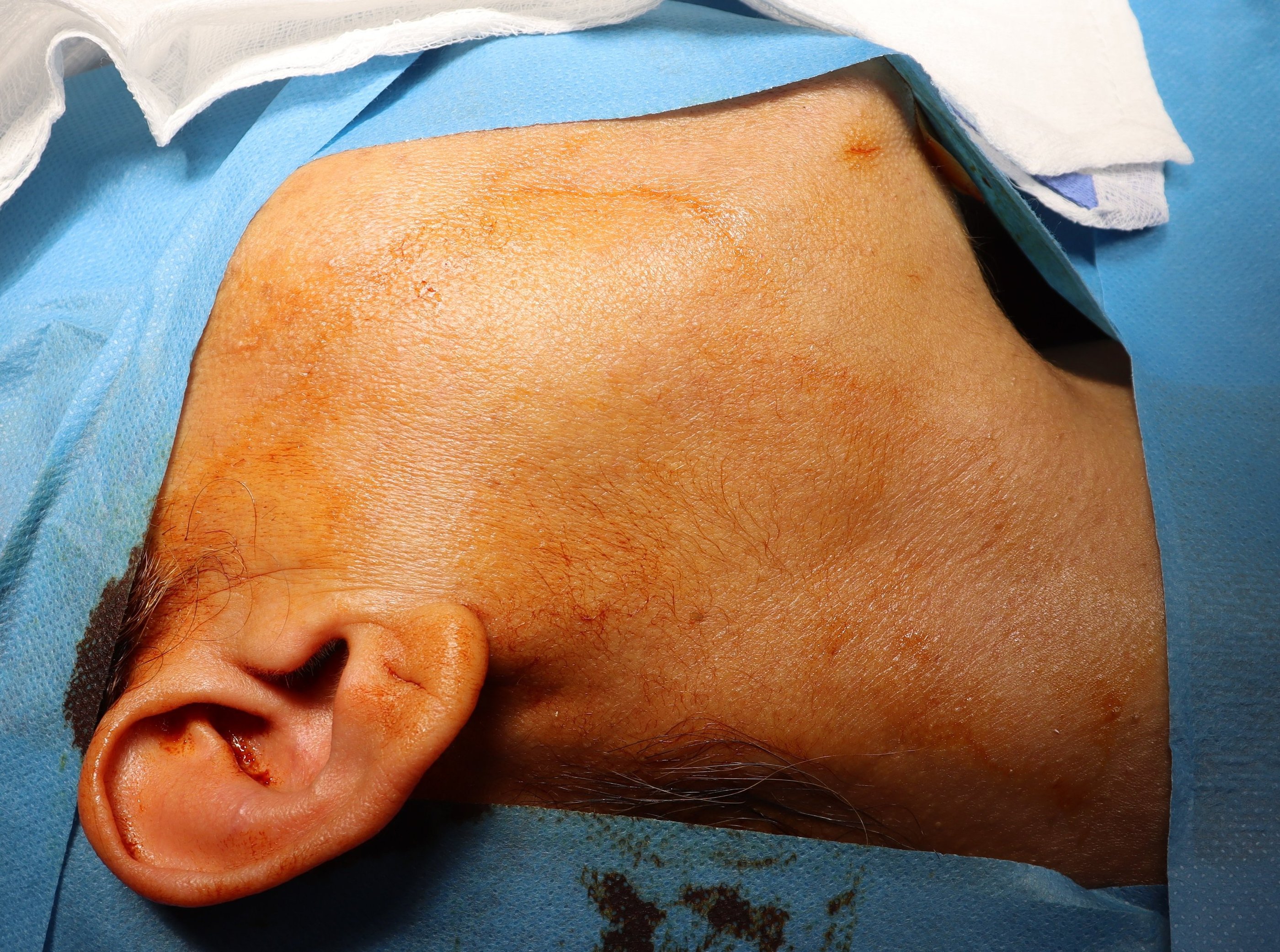
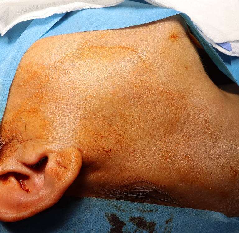
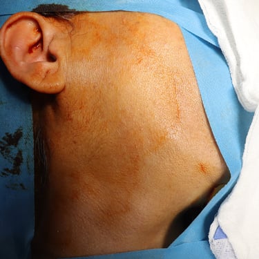
A 51-year-old female presented with a one-year history of a slowly growing, painless swelling in the right parotid region. She is a non-smoker with no significant past medical, surgical, or drug history. On physical examination, she was classified as G1 (indicating a clinically mild glandular involvement).
SURGERYHEAD AND NECK
4/5/20252 min read
Patient Information:
A 51-year-old female presented with a one-year history of a slowly growing, painless swelling in the right parotid region. She is a non-smoker with no significant past medical, surgical, or drug history. On physical examination, she was classified as G1 (indicating a clinically mild glandular involvement).
Laboratory Investigations:
Thyroid function tests showed a mildly elevated TSH (6.59 µIU/mL) with a normal free T4 (14.3 pmol/L), suggesting subclinical hypothyroidism. The anti-thyroid peroxidase antibody (ATPO) level was elevated (247 IU/mL), indicating an underlying autoimmune thyroid condition, possibly Hashimoto’s thyroiditis.
Neck and Parotid Ultrasound Findings:
Thyroid Gland:
The right lobe was mildly enlarged (65 × 28 × 24 mm), while the left lobe was normal in size (55 × 17 × 15 mm).
The gland showed an inhomogeneous echotexture, with multiple well-defined nodules.
The largest nodule in the right lobe measured 26 × 18 × 17 mm (TR2), while the largest in the left lobe measured 12 × 9 × 8 mm (TR3).
Mild increased vascularity was noted without microcalcifications or retrosternal extension.
No significant cervical lymphadenopathy was detected.
Salivary Glands:
The parotid and submandibular glands appeared normal, except for a small complex nodule (16 × 14 × 11 mm) in the superficial lobe of the right parotid gland.
The lesion had increased in size compared to a prior ultrasound, prompting further evaluation.
Fine Needle Aspiration Cytology (FNAC):
A fine-needle aspiration biopsy (FNAB) of the right parotid gland nodule was performed, and cytology findings were consistent with a benign neoplasm (Category IVA of the Milan system), suggestive of a pleomorphic adenoma (benign mixed tumor of the salivary gland).
Management Plan:
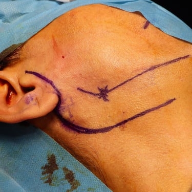
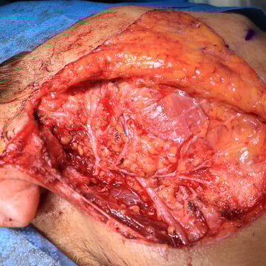
Given the increasing size of the right parotid gland nodule and the risk of malignant transformation in pleomorphic adenomas, the patient was scheduled for a right superficial parotidectomy. Additionally, a scalp cyst excision was planned as an incidental procedure.
Surgical Outcome and Histopathology:
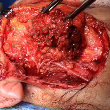
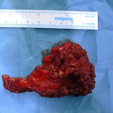
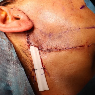
Right Superficial Parotidectomy:
Histopathology confirmed pleomorphic adenoma, with no evidence of malignancy.
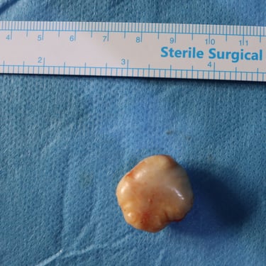
Scalp Cyst Excision:
The histopathology identified a pilar (trichilemmal) cyst, with no malignant features.
Follow-Up and Prognosis:
The patient had an uneventful postoperative recovery. Long-term follow-up was advised due to the potential for pleomorphic adenoma recurrence, although the surgical margins were clear. Endocrine follow-up was also recommended to monitor thyroid function and manage subclinical hypothyroidism, particularly given the presence of positive ATPO antibodies.
Gallery
Address
Majid Bag Main Street, Beside University of Sulaymaniyah Old Campus, Madam Mitterrand, Sulaymaniyah, Iraq
Contacts
info@ssthyroid.com