Spindle Cell Sarcoma in an 86-Year-Old Male
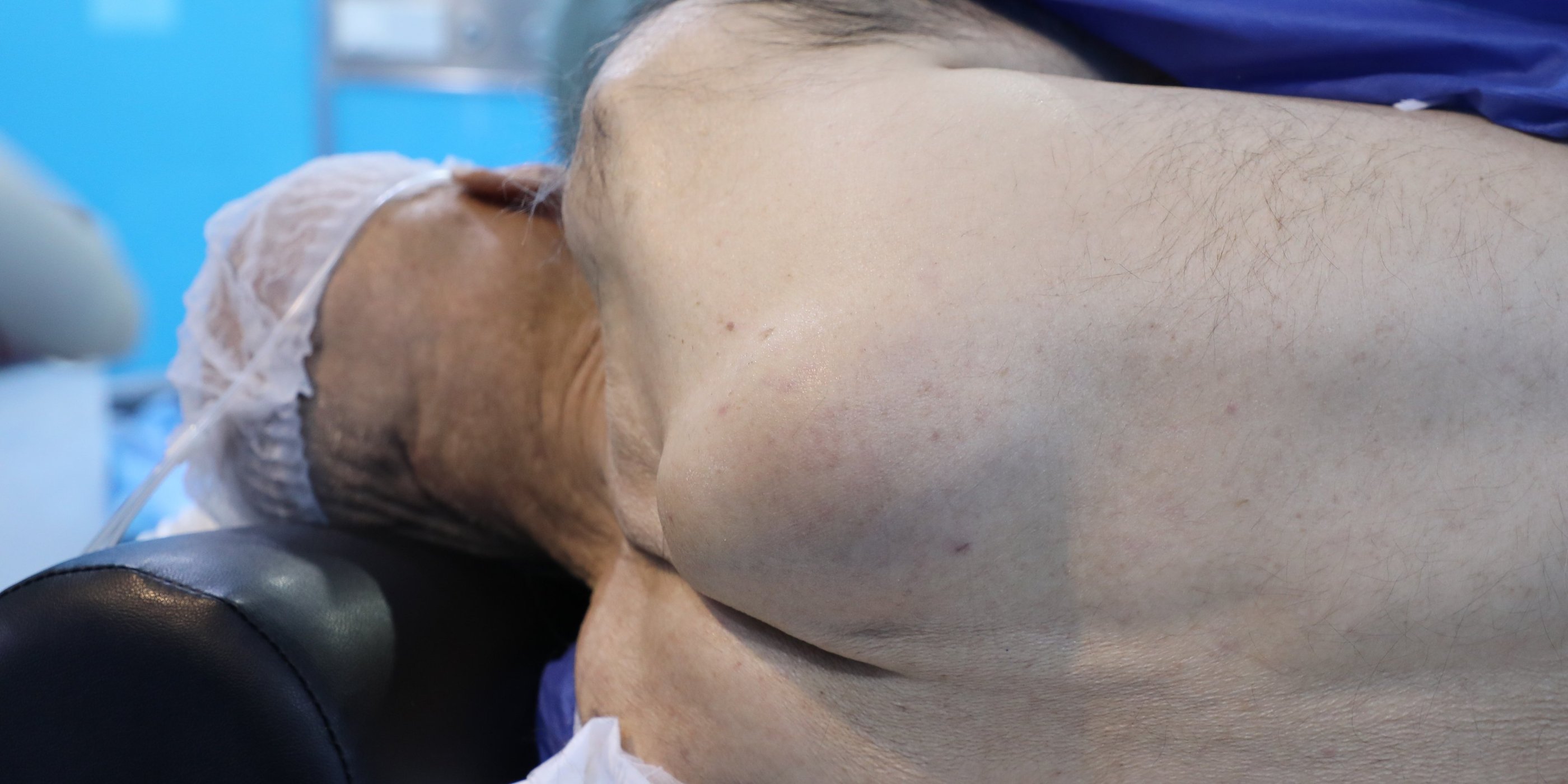
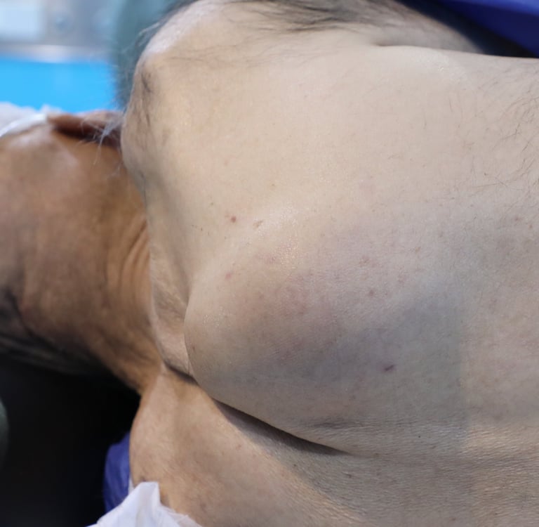
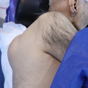
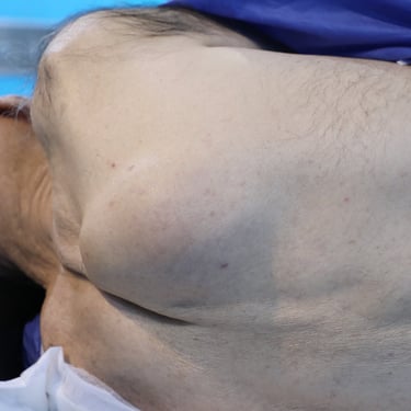
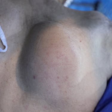
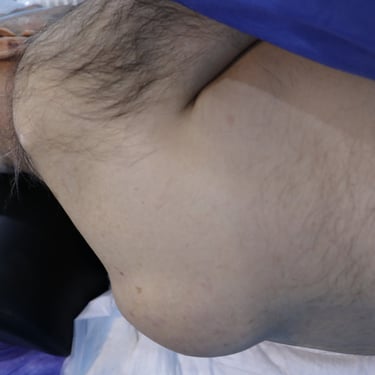
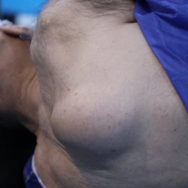
An 86-year-old male presented with a progressively enlarging back mass for the past two months. He was a non-smoker. His past medical history was significant for hypertension (HTN), ischemic heart disease (IHD), and percutaneous coronary intervention (PCI). Additionally, he had undergone femur surgery in the past.
SURGERY
4/12/20251 min read
Patient Information:
An 86-year-old male presented with a progressively enlarging back mass for the past two months. He was a non-smoker. His past medical history was significant for hypertension (HTN), ischemic heart disease (IHD), and percutaneous coronary intervention (PCI). Additionally, he had undergone femur surgery in the past.
Laboratory Investigations:
Thyroid function tests revealed a mildly suppressed thyroid-stimulating hormone (TSH) level of 0.108 µIU/mL, while free thyroxine (FT4) was within normal limits at 19.8 pmol/L.
Chest CT with IV Contrast:
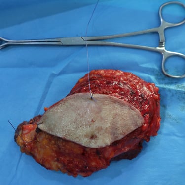
A contrast-enhanced CT scan of the chest was performed to evaluate the posterior back mass. Imaging revealed a well-defined soft tissue density mass located in the right upper back, measuring 8 × 4.5 cm. The lesion was situated in the subcutaneous fat, superficial to and inseparable from the right trapezius muscle. No internal fat or calcification was noted. The mass exhibited mild heterogeneous post-contrast enhancement on the venous phase. The underlying scapula appeared normal. The radiological impression was suggestive of a soft tissue sarcoma, and tissue diagnosis was recommended.
Core Needle Biopsy:
Under local anesthesia and ultrasound guidance, seven Tru-Cut core biopsies were obtained from the large posterior chest wall mass. Histopathological examination identified the lesion as a spindle cell sarcoma, and further immunohistochemical staining was advised.
Histopathology Report:
The biopsy revealed a spindle cell sarcoma with a mitotic rate of 19 per 2mm², indicating a high proliferative index. No necrosis or lymphovascular invasion was identified. Immunohistochemical analysis was recommended to confirm the diagnosis, including markers such as SS18-SSX, CD99, MUC4, S100, SMA, Desmin, STAT6, and CD34, with the possibility of additional markers if needed. The primary suspicion was monophasic synovial sarcoma, pending further immunostaining for definitive characterization.
Gallery
Address
Majid Bag Main Street, Beside University of Sulaymaniyah Old Campus, Madam Mitterrand, Sulaymaniyah, Iraq
Contacts
info@ssthyroid.com